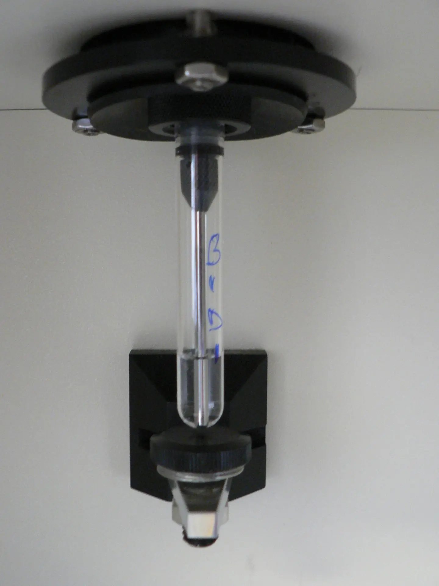
Flow Cytometry: An Advanced Tool for Rapid and Sensitive Detection of MAP Infection
Introduction to Flow Cytometry
An advanced laboratory method for examining the physical and chemical properties of cells or particles suspended in a fluid stream, flow cytometry (FC) is used extensively in immunology, microbiology, and veterinary diagnostics. In order to detect Mycobacterium avium subspecies paratuberculosis (MAP), flow cytometry uses the interaction between intact MAP bacteria and antibodies found in the serum of infected animals. This enables a quick, accurate, and early diagnosis of Johne's disease, a chronic intestinal infection that significantly reduces livestock industry profits.
Principle of Flow Cytometry for MAP Detection
In the FC assay, intact MAP cells serve as the target particle that travels through the flow cytometer as well as the test antigen. By guaranteeing that the entire repertoire of unaltered MAP surface proteins is accessible for antibody binding, this technique enables the detection of particular immune responses directed against the bacterium.
Whole MAP bacteria are incubated with serum samples from suspected animals.
The bacterial surface antigens are bound by MAP-specific antibodies.
Fluorescent secondary antibodies are used to identify these antibody-bound bacteria.
The fluorescence intensity of each bacterium is measured as it moves through a laser beam inside the flow cytometer.
The information gathered enables the measurement of serum antibodies specific to MAP, a sign of infection.
Advantages of Flow Cytometry in MAP Diagnosis
1. Speed and Efficiency
Flow cytometry produces results in less than four hours, in contrast to traditional culture techniques that take weeks (up to 12–16 weeks) to grow MAP colonies. Farmers and veterinarians can make decisions about infection control and herd management more quickly thanks to this impressive reduction in diagnostic time.
2. High Sensitivity and Specificity
The reliability of FC assays in identifying MAP infection has been shown by experimental research:
- Approximately 95% sensitivity shows that the assay can accurately identify animals that are infected.
- With a specificity of roughly 97%, it can accurately rule out animals that are not infected.
- The detection of IgG1 antibodies specific to MAP in adult cattle achieved a perfect specificity of 100% and a sensitivity of 78%.
This degree of accuracy validates FC as a trustworthy diagnostic substitute and outperforms many conventional serological techniques.
3. Early Detection Capability
Finding infected animals before clinical symptoms show up is one of the most important challenges in managing Johne's disease. As early as 170 days after infection, the FC assay can identify MAP-specific antibodies in calves, allowing for early intervention and lowering the risk of transmission within herds.
Practical Applications of Flow Cytometry in MAP Research and Veterinary Medicine
Herd Screening and Surveillance
Large-scale screening programs benefit from FC assays, which assist in identifying infected animals in subclinical stages so that prompt control measures, like culling or segregation, can be put in place.
Epidemiological Investigations
Researchers can investigate the dynamics of MAP transmission both within and between herds, identify risk factors, and assess the efficacy of biosecurity measures by tracking antibody profiles.
Evaluation of Immune Responses
In order to support the creation of novel control strategies, flow cytometry plays a crucial role in tracking the immune response to experimental vaccines or therapeutic interventions.
Complementary Tool in Diagnostic Panels
FC is frequently used in conjunction with fecal culture, PCR, and ELISA tests to increase overall diagnostic accuracy because of its high specificity and early detection capability.

