Fluorescent Microscopy and Fluorescent Antibody Test (FAT) for Detection of MAP
Introduction
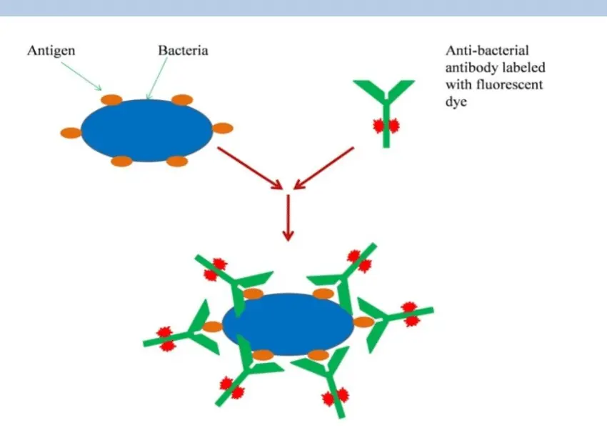
Fluorescent microscopy, when applied in the Fluorescent Antibody Test (FAT), provides a powerful technique for the detection and localization of pathogens such as Mycobacterium avium subsp. paratuberculosis (MAP), especially in biological specimens like milk, tissue, or smears. FAT relies on the use of fluorescent-labeled antibodies that specifically bind to MAP antigens. Upon binding, the labeled antibodies emit fluorescence when excited by a specific wavelength of light under a fluorescence microscope. This principle enables direct visualization of the bacteria in situ, providing both qualitative and semi-quantitative results.
Principle of the Fluorescent Antibody Test
The FAT is based on immunofluorescence—a method that uses antibodies tagged with fluorogenic molecules such as fluorescein isothiocyanate (FITC). The general steps involved in the test include:
- Sample Preparation: Samples such as pasteurized milk, vaginal smears, or tissue sections are fixed onto slides.
- Application of Labeled Antibody: A MAP-specific antibody, conjugated with a fluorophore, is applied to the fixed specimen.
- Incubation and Washing: After allowing time for binding, unbound antibodies are washed away.
- Microscopy: The slides are examined under a fluorescence microscope equipped with filters that excite the fluorophore, revealing bright fluorescent signals where antigen-antibody complexes have formed.
One of the key features of this method is its use of enzymatically activated fluorophores. For example, the fluorigenic substrate may be converted into carboxyfluorescein in live cells, enhancing fluorescence signal and aiding in the visualization of viable MAP bacilli.
Applications of FAT in MAP Detection
The FAT has been successfully employed to detect low levels of MAP (as few as ∼10² colony-forming units per milliliter) in various sample types:
- Pasteurized Milk: D’Haese et al. (2003) demonstrated a sensitivity of approximately 73% when using FAT to detect MAP in pasteurized milk, a matrix where bacterial numbers may be low and viability is often reduced.
- Raw Milk and Vaginal Smears: FAT has also proven valuable in screening vaginal smears and raw milk, especially in herds known to be infected or in animals showing early clinical signs.
- Tissue Specimens: FAT enables rapid identification of MAP bacilli in tissue biopsies, such as intestinal mucosa and mesenteric lymph nodes, even in subclinical or early-stage infections.
Advantages of FAT
- Rapid Turnaround: Results can be obtained within a few hours, much faster than culture-based methods that may take weeks.
- High Specificity: Gilmour and Angus (1976) reported that the FAT provided a specific immune response in cattle experimentally infected with M. avium subsp. avium and M. avium subsp. paratuberculosis.
- Detection in Complex Matrices: The FAT performs well even in complex samples like milk or mucosal smears, where other techniques may be hindered by inhibitors or sample degradation.
- Viable Cell Detection: Due to the enzymatic activation of fluorophores, this method may favor detection of live, metabolically active MAP cells, a critical aspect in assessing infectious risk.
Innovations and Enhancements
Goudswaard et al. (1977) proposed the incorporation of the Defined Antigen Substrate Spheres (DASS) system to improve specificity and enable routine use of FAT for antibody detection against Mycobacterium species. The DASS platform standardizes antigen presentation, reducing variability and enhancing the reproducibility of results.
Moreover, advancements in fluorescent dyes and image analysis software are allowing more quantitative and automated interpretations of FAT results, making it more feasible for high-throughput screening and surveillance programs.
Diagnostic Role and Research Use
FAT plays a vital role in confirmatory diagnosis of MAP infections, especially in cases where rapid answers are needed or where other methods yield inconclusive results. In research contexts, it serves as an important tool for:
- Studying infection dynamics in tissues
- Monitoring MAP shedding in milk
- Evaluating vaccine efficacy by assessing antibody response
- Supporting epidemiological surveys in livestock populations
Conclusion
The Fluorescent Antibody Test (FAT) is a valuable diagnostic method for the rapid, specific detection of Mycobacterium avium subsp. paratuberculosis in a range of clinical samples. With a sensitivity of approximately 73% in pasteurized milk and the ability to detect low concentrations of MAP, FAT serves as an effective screening and confirmation tool, particularly when rapid diagnosis is required. Continued refinements in fluorophore chemistry and antibody development will further improve its diagnostic potential, making it an integral component in the detection and management of paratuberculosis in domestic animals.
