Culture-Based Diagnostic Test for MAP Detection
Introduction
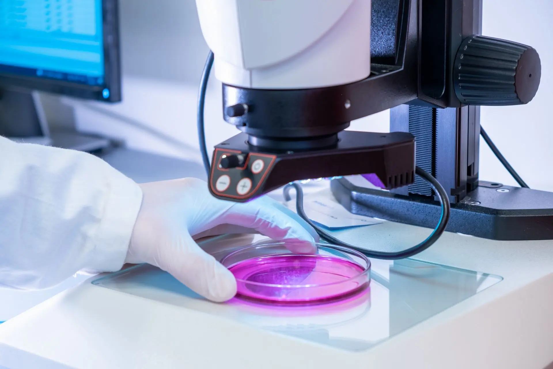
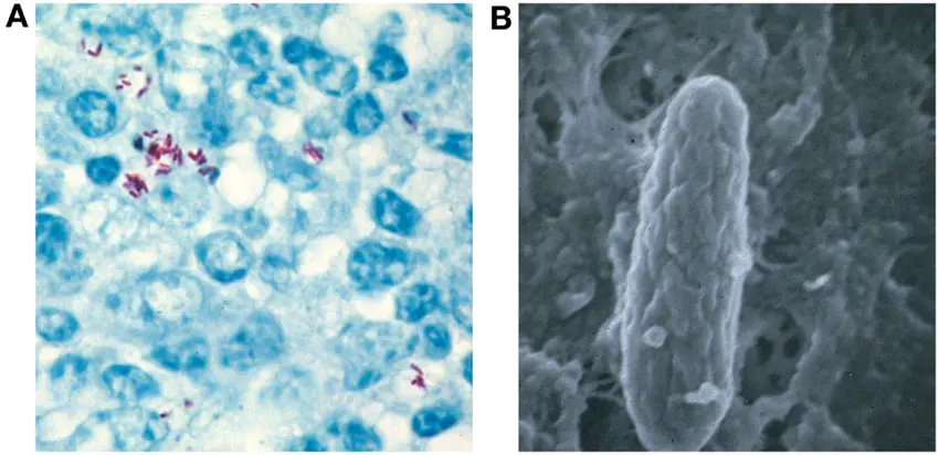
The culture of Mycobacterium avium subsp. paratuberculosis (MAP) remains the gold standard for confirming MAP infection due to its ability to detect viable bacteria. Despite being time-consuming and technically demanding, culture is the most definitive method for diagnosing paratuberculosis (Johne's disease) in animals, particularly in ruminants. This method is central to surveillance, research, and control programs targeting MAP.
Sample Sources and Relevance
MAP can be isolated from a wide range of biological and environmental samples, depending on the infection stage and the diagnostic purpose. Common sources include:
- Faeces: Frequently used for screening live animals; however, due to intermittent shedding, multiple samples are often required.
- Milk: Especially in dairy animals, milk samples are used to detect shedding through the mammary gland.
- Tissue: Samples from ileum, mesenteric lymph nodes, or liver collected during necropsy offer a high yield, particularly in clinically infected animals.
- Blood: While not routinely used for culturing MAP, blood can occasionally be tested.
- Environmental samples: Manure, soil, and water samples from contaminated premises are used to assess herd contamination or farm-level infection status.
These samples help determine not only individual infection status but also environmental contamination, which plays a key role in transmission.
Culture Conditions and Duration
MAP is a slow-growing organism, making culture a long and sometimes unpredictable process. Two primary types of media are used:
-
Solid media: Such as Herrold’s Egg Yolk Medium (HEYM) supplemented with mycobactin J.
- Colonies typically appear in 2–4 months, although growth may extend up to 6–8 months, especially in samples with low bacterial load.
- Colonies are small, round, and non-pigmented with rough surfaces.
-
Liquid media: Such as Middlebrook 7H9 broth or automated liquid systems (e.g., BACTEC MGIT 960).
- Offers faster results, often within 6–7 weeks.
- However, the extended incubation needed increases the risk of contamination, especially from faster-growing environmental mycobacteria or fungi.
Advances in Culture Technology
To overcome the lengthy duration of traditional culture, automated culture systems have been developed:
-
BACTEC MGIT 960 System:
- Reduces detection time to 4–7 weeks.
- Can detect as low as 10 colony-forming units (CFU)/ml (Shin et al., 2007).
- Uses fluorescence-based oxygen consumption as an indicator of bacterial growth.
- Other semi-automated systems and methods like TiKa MAP culture and phage-based detection are under development to accelerate MAP isolation while maintaining specificity and sensitivity.
Diagnostic Relevance
Culture results provide definitive evidence of infection, especially when colony morphology and mycobactin dependence are considered.
However, interpretation of culture results must consider:
- Intermittent shedding in faeces, which means serial sampling is recommended for a reliable diagnosis.
- False negatives may occur in early infections or subclinical animals due to low bacterial load.
- The high cost and time demand, which make it unsuitable for large-scale rapid screening.
Advantages of Culture
- Definitive Diagnosis: Confirms the presence of viable MAP organisms.
- Phenotypic Characterization: Enables strain typing, antibiotic susceptibility testing, and genotyping.
- High Specificity: Especially when used in conjunction with molecular confirmation (e.g., PCR targeting IS900).
-
Useful for Surveillance: Helps in assessing contamination levels in herd environments and guiding biosecurity measures.
🧠 Conclusion
Although slow and resource-intensive, culture remains the cornerstone of MAP detection in veterinary diagnostics. Its ability to isolate live organisms makes it crucial for confirmatory testing, epidemiological investigations, and research studies. Innovations such as automated culture systems and improved decontamination protocols are helping to overcome some of the limitations associated with traditional culture. However, for routine diagnostics and herd screening, culture is often used in combination with molecular tools (PCR) and serological tests (ELISA) to achieve a more comprehensive and time-efficient diagnosis.
